
Brachycephalic breeds are predisposed to fluid accumulation in the Tympanic Bulla (TB). Middle ear fluid can be sterile effusion or may be secondarily infected with evidence of microbial overgrowth on cytology and/or culture. Affected dogs may be symptomatic or asymptomatic. Middle ear effusion may be an incidental finding in many brachycephalics, which complicates the diagnosis of Otitis Media (OM) in these breeds.
Additionally, the brachycephalic external ear canal exhibits extreme narrowing (stenosis) compared to dolichocephalic and mesocephalic breeds, which predisposes them to otitis and secondary infection.
EMERGING EVIDENCE
- Paterson S. Otitis media with effusion in the boxer: a report of seven cases. J Small Anim Pract. 2017 Dec 12. doi: 10.1111/jsap.12801. Epub ahead of print. PMID: 29231979.
- Töpfer T, Köhler C, Rösch S, Oechtering G. Brachycephaly in French bulldogs and pugs is associated with narrow ear canals. Vet Dermatol. 2022 Jun;33(3):214-e60. doi: 10.1111/vde.13067. Epub 2022 Mar 16. PMID: 35293639.
- Milne E, Nuttall T, Marioni-Henry K, Piccinelli C, Schwarz T, Azar A, Harris J, Duncan J, Cheeseman M. Cytological and microbiological characteristics of middle ear effusions in brachycephalic dogs. J Vet Intern Med. 2020 Jul;34(4):1454-1463. doi: 10.1111/jvim.15792. Epub 2020 May 14. PMID: 32407559; PMCID: PMC7379010
CLINICAL PRESENTATION
Clinical signs vary greatly amongst affected individuals. It is not uncommon for dogs to present with otic pruritus, erythema and otic discharge, without any evidence of middle ear involvement. Others can present with acute onset vestibular and/or neurological clinical signs, including but not limited to; head tilt to the affected side, nystagmus, facial palsy or paralysis, vestibular anomalies, neurogenic KCS, reduced or absent hearing, dysphagia, hypersalivation, lethargy, inappetence etc. Oftentimes, there is a history of otitis externa or allergic dermatitis, but this is not always the case.
DIAGNOSTIC APPROACH
- Computed Tomography (CT): The gold standard imaging modality used for assessment of the TB and associated bony and soft tissue structures. Oftentimes, intravenous contrast is administered to aid visualisation of the affected area, and to identify structures with a collateral blood supply. Contrast is useful in differentiating between middle ear effusion and a middle ear mass (tumours, cholesteatoma, etc). Radiography nowadays is an insensitive imaging technique for investigation of OM and is no longer recommended. Instead, patients should be referred to a facility with a CT.
- Magnetic Resonance Imaging (MRI): Less sensitive than CT for assessing the tympanic bulla. However, a Specialist Radiologist can usually interpret the findings of an MRI sufficiently, such that a CT is not required as an additional diagnostic test. MRI is useful for evaluation of the brain and soft tissues, which is helpful for investigation of other differential diagnoses such as a brain tumour or ischaemic event that may have caused the presenting clinical signs.
- Otoscopy: If an intact tympanic membrane (TM) is visualised on otoscopic exam, this does not rule out otitis media. A healthy canine TM should heal within 6-8 weeks, meaning a prior perforation may not be appreciated if sufficient time has passed. Similarly, although a bulging TM is often thought to indicate OM, a non-bulging TM does not rule out otitis media. More often than not, the TM in brachycephalic dogs cannot be completely visualised due to breed related stenosis. Ceruminoliths are commonly found in the external ear canals of dogs with otitis, which also contribute to poor TM visualisation.
- Cytology: Of the external and middle ear contents should be performed in every patient.
- Microbial Culture and Sensitivity of the Middle Ear: should be performed in every patient (see below).
TREATMENT
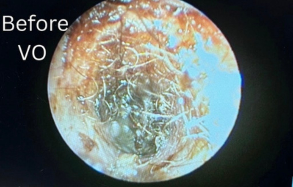
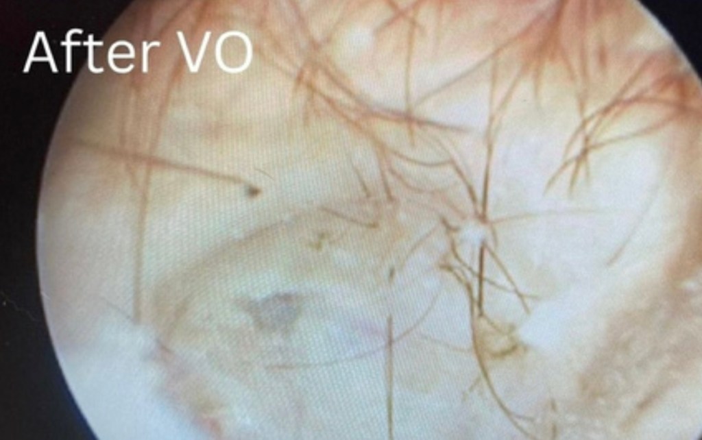
If Otitis Media is identified, and the dog is symptomatic, the patient should be referred to a Veterinary Dermatologist for ongoing care and management. If left untreated, otitis media can progress to otitis interna. Video Otoscopy (VO): The preferred technique to visualise and clean the external ear canal, determine whether the TM is perforated or intact, collect biopsies of abnormal tissue and/or masses and perform a myringotomy if required. A myringotomy (intentional, surgical incision into the caudoventral portion of the pars tensa of the TM) is performed under general anaesthesia to gain access to the TB. Middle ear effusion should be carefully collected and submitted to an external laboratory for bacterial and fungal culture and sensitivity. Cytological examination of the fluid should also be performed. Following myringotomy, the TB is thoroughly and repeatedly flushed using warm, sterile 0.9% NaCl via the working channel of the VO. The bullae and external canal are then aspirated to remove all remaining fluid and debris, and to ensure the ear is left clean and dry.
- Systemic Antimicrobials are Indicated in Cases of OM: Antibiotic choice should be guided by microbial culture and sensitivity results. Empirical antibiotic treatment with Clavulox (22g/kg BID) or clindamycin (11mg/kg BID) should be prescribed post-operatively, whilst awaiting culture results. Antibiotics are continued for 6-8 weeks total.
- Systemic Corticosteroids: Are the mainstay of treatment for OM in human medicine. In dogs, 1mg/kg (tapering) is recommended post myringotomy for anti-inflammatory and analgesic benefits.
- Topical Otic Therapy: Should also be guided by microbial culture & sensitivity. Ear drops instilled post myringotomy must be middle ear safe in order to prevent ototoxicity and potential deafness. Ear drops should contain a topical steroid, such as 0.2% dexamethasone.
- Severe OM: Cases that are chronic, recurrent, non-responsive to medical treatment, neoplastic etc. should be considered end stage ear disease. These dogs should be referred to a Specialist Veterinary Surgeon for TECALBO (Total Ear Canal Ablation Lateral Bulla Osteotomy) to prevent ongoing pain and suffering.
KEY TAKE AWAYS
Otitis in brachycephalic breeds is complex in nature. Affected animals should ideally be referred to a multidisciplinary specialist hospital for assessment by dermatologists, radiologists, neurologists and surgeons where necessary for treatment and ongoing care.