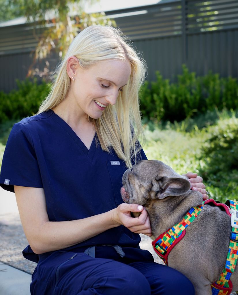

Intertriginous dermatitis (aka intertrigo) refers to inflammation and/or infection of any skin fold on the body e.g. lip folds (cheilitis), axillae, peri-vulvar, tail fold, interdigital etc. The underlying cause of intertriginous dermatitis is a result of both mechanical trauma (friction due to skin and hair rubbing) and optimal conditions for microbes to flourish, such as warmth, humidity and moisture. Excretions such as tears and saliva, as well as sebum, glandular secretions and overzealous cleaning of the skin causes inflammation. This leads to microbial dysbiosis and subsequent secondary Malassezia and/or bacterial infection, resulting in a perpetuating cycle of disease.
In brachycephalic breeds, intertriginous dermatitis of the nasomaxillary skin fold is a common presentation. Their unique anatomy, concomitant ophthalmic and/or dermatological issues, make brachycephalics predisposed to intertriginous dermatitis. This can be a challenging condition for veterinarians to treat given the multiple underlying causes, high recurrence rate and thus, need for ongoing management.
EMERGING EVIDENCE
- Rexo A, Hansen B, Clarsund M, Krumbeck JA, Bernstein J. Effect of topical medication on the nasomaxillary skin-fold microbiome in French bulldogs. Vet Dermatol. 2022 Feb;33(1):10-e5. doi: 10.1111/vde.13017. Epub 2021 Oct 19. PMID: 34668256.
- O’NeillI, D.G., Rowe, D., Brodbelt, D.C. et al. Ironing out the wrinkles and folds in the epidemiology of skin fold dermatitis in dog breeds in the UK. Sci Rep 12, 10553 (2022).
CLINICAL PRESENTATION
Intertriginous dermatitis may present with erythema, crusting, keratosebaceous debris, alopecia, malodour and pruritus. In severe and/or chronic cases, pain, ulceration, lichenification and hyperpigmentation can occur. Often, a combination of clinical signs are present.
DIAGNOSTIC APPROACH
- Cytology: Cytological evaluation of the affected area of skin is paramount for treatment guidance. Sticky tape prep is the preferred cytology collection technique due to malleability of the tape between delicate skin fold areas. The affected skin may need to be gently dried with a tissue prior to cytological collection to facilitate the tape sticking to the skin surface.
- Rule out demodicosis: Demodex mites are considered commensal microfauna on canine skin and usually exist in low numbers on healthy canine skin. Demodicosis occurs when mites opportunistically increase in number when there is local inflammation and immunocompromise. Ensure ectoparasite prophylaxis is current, with products such as Nexgard, Simparica or Bravecto.
- Dermatohistopathology: Rarely, tissue biopsies may be required for histopathological evaluation. This may be necessary in non-responsive cases, or in cases where deep pyoderma is present.
TREATMENT
According to the 2025 Antimicrobial use guidelines for canine pyoderma by the International Society for Companion Animal Infectious Diseases (ISCAID) (Loeffler et.al), topical therapy should be considered as the sole, first-line treatment for superficial pyoderma cases, such as intertriginous dermatitis. Furthermore, topical antiseptics should be favoured over topical antibiotics, due to the rise of multi-drug resistance (MDR) bacteria.
Preferred Topical Treatments Include:
- Chlorhexidine 2-4%
- Silver sulfadiazine 1% cream
- Topical otic preparations (gels, suspensions, lotions) containing an antibiotic, anti-fungal and steroid.
- If rods are present on cytology, consider using a topical product containing gentamicin or a fluoroquinolone such as marbofloxacin or enrofloxacin.
- Topical ophthalmic preparations (drops or ointments) containing and antibiotic and steroid.
- These are useful if the affected skin is located close to the globe.
- Topical steroids (ointments, creams, lotions) e.g. mometasone furoate, betamethasone dipropionate, hydrocortisone aceponate.
Occasionally, systemic anti-microbials are needed, and should be assessed on a case-by-case basis.
A short, tapering course of oral prednisolone (0.5-1mg/kg; 7-14days) can be useful to address associated inflammation, pain and pruritus.
Weight loss is encouraged in obese animals as accumulation of subcutaneous fat will contribute to skin fold dermatitis.
Surgical resection of the apposing skin surfaces should be considered in refractory or severe cases whereby medical management proves ineffective or is unsustainable for the pet and/or owner.
MAINTENANCE AND PREVENTION
Successful management requires identification and treatment of the underlying disease.
An example of a topical maintenance regime for brachycephalic nasal folds is 2-4% chlorhexidine wipes followed by topical mometasone (Elocon cream; Zatamil gel). This should be repeated twice weekly.
KEY TAKE AWAYS
- Overcleaning skin folds, excessive moisture and mechanical friction can cause inflammation, microbial dysbiosis and can lead to secondary infection.
- Underlying issues such as allergic dermatitis, ophthalmic disease etc. must be concurrently addressed in order to successfully manage nasomaxillary intertrigo in brachycephalic breeds.
- Intertriginous dermatitis requires ongoing management tailored to the individual pet and owner.